- SEIKA Digital Image Corporation.
- ProductList
- PIV(Particle Image Velocimetry)
- Micro-area fluid measurement Micro PIV/Micro LIF
Micro-area fluid measurement Micro PIV/Micro LIF
Micro PIV/LIF System
Micro PIV and micro LIF are systems for analyzing the flow velocity distribution, mixing, and diffusion of microfluidics (microfluidics). A system is constructed by combining optical systems such as a microscope and components for micro PIV such as dedicated tracer particles. It is widely used in the development of micro biochemical chips called micro TAS and lab-on-chip in recent years.
Some links lead to Japanese pages.
Feature
- Various latest micro-PIV analysis techniques developed for micro-PIV such as average correlation and SAT-PTV are abundantly incorporated.
- We have a lineup of various types of micro PIV systems such as 2D micro PIV, 3D micro PIV, and confocal micro PIV, and support the research and development of microfluidics technology.
- SAT-PIV Algorithm
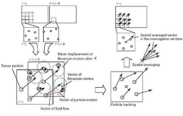
The SAT-PTV algorithm (*1) is a velocity analysis algorithm that can greatly reduce the effects of Brownian motion. Brownian motion is the most important error factor in micro-PIV. Seika Digital Image's PIV analysis software, Koncerto II, incorporates an analysis algorithm specialized for micro PIV to achieve highly accurate analysis. Learn more about the SAT-PIV algorithm here.
Application
- μTAS
- Lab on a chip microfluidics
- piezo element
- MEMS・BioMEMS
- micro chemical process
System list
Micro PIV System
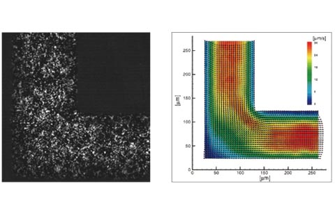
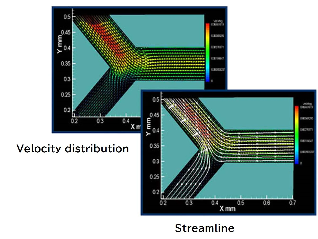
Flow Velocity Distribution in a 100 μm Microchannel Using Confocal Scanning Micro-PIV
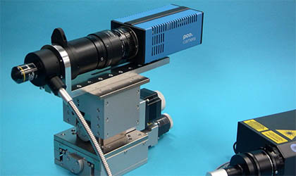
In addition to conventional double pulse type micro PIV (2D, stereo 3D), time series micro PIV, we have a lineup of various types of micro PIV systems such as confocal scanning micro PIV. The dedicated micro-optical system has a lineup of models that are compatible with high-power pulse lasers and UV lasers. Peripheral equipment such as a focus scanner that can scan the focal plane at high speed synchronously with the high-speed video frame is also available. We also have a wide variety of fluorescent particles, which are indispensable for micro PIV, that are optimal for tracers, and can be selected according to color, size, and target fluid (water, organic solvent, etc.).
2D-micro PIV system (double pulse type)
■ Double shutter type PIV camera
■ Double pulse laser 30mJ/pulse 15Hz 532nm
■ Timing controller
■ Micro optical system, fiber bundle delivery type
■ Control analysis software Koncerto II micro
■ PC for control analysis
3D-micro PIV system (double pulse type)
■ Double shutter type PIV camera x 2
■ Double pulse laser 30mJ/pulse 15Hz 532nm
■ Timing controller
■ Micro stereo optical system fiber bundle delivery type
■ Control analysis software KoncertoⅡmicro 3D
■ PC for control analysis
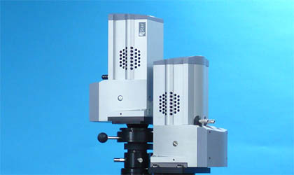
Micro LIF system
Data provided by: Keio University Hishida/Sato Laboratory
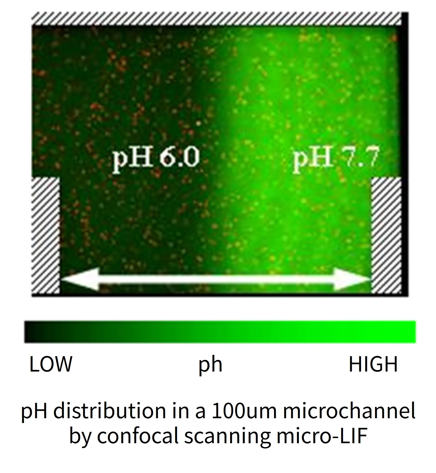
Micro LIF can measure concentration distribution, temperature distribution, diffusion, mixing, reaction, pH, etc. The dedicated micro-optical system can co-axially illuminate a high-power UV pulse laser, which is not possible with general microscopes.
System configuration example
■Double shutter type LIF camera
■Double pulse laser 30mJ/pulse 15Hz 532nm
■Timing controller
■Micro optical system, fiber bundle delivery type
■Control analysis software Koncerto II LIF
■PC for control analysis
Confocal micro PIV/micro LIF system
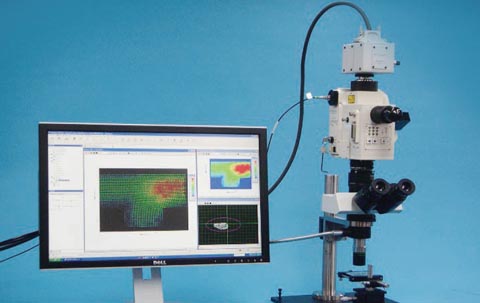
Confocal micro PIV/LIF is a system that enables high-precision measurement by increasing the resolution in the Z-axis direction by using a confocal scanner in addition to the optical system of a normal microscope.
Why is confocal imaging effective in microimaging?
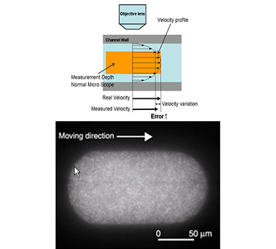
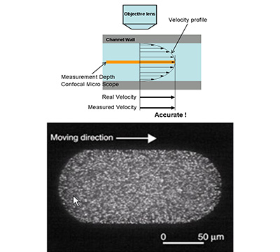
Micro PIV uses the concept of measurement depth (MD) instead of depth of field (DOF) when defining Z-direction resolution.
This is a concept proposed by Minehart et al., UC Santa Barbara, and refers to the range in which the light intensity of the particle image appears strong enough to affect velocity measurements, and is generally much thicker than the DOF. In a normal micro PIV, the MD is much thicker than the designed depth of focus of the objective lens, and different velocity components are mixed within the thickness of the MD, resulting in measurement errors. (see figure), on the other hand, in confocal scanning micro PIV, it is possible to make the MD thin so that only a single velocity component is included in the MD area, enabling high-precision measurement distribution in microfluidics. rice field.
Micro LIF usually does not use fluorescent particles, but rather uses a fluorescent agent dissolved in a liquid, and the liquid itself emits light. Therefore, in normal microscope observation, even if a high-NA objective lens is used, fluorescence emission in front of and behind the focal plane enters as stray light, lowering the spatial resolution in the optical axis direction (Z direction). (The spatial resolution in the XY direction is significantly lower than the spatial resolution in the Z direction.) With confocal imaging, the fluorescence emission in front of and behind the focal plane can be almost completely cut off, resulting in a high Z-direction resolution that matches the XY-direction resolution. It can be obtained.
System configuration example
■ Ultra-sensitive high-speed video camera
■ Confocal scanner CSU-X1
■ CW DPSS Laser 100mW 488nm
■ Focus scanner FS-100 (option)
■ Micro optical system UFS-200 Fiber bundle delivery type
■ Control analysis software Koncerto II
■ PC for control analysis
Brightfield image
Confocal image
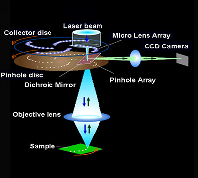
Principle of confocal scanner
Confocal scanner
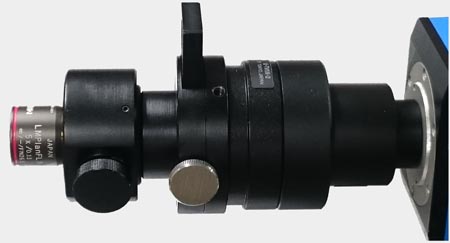
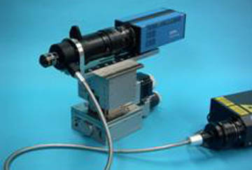
Optical system for micro imaging
UFS-200 UFS-500
The optical system dedicated to micro imaging is compatible with high-power pulse lasers, UV lasers, etc.
This is a flexible microscope system that can be installed at any angle, such as sideways or obliquely, depending on the specimen.
UFS-200
A compact type featuring a highly efficient dichroic cube.
The distance between the dichroic mirror for laser light introduction and the objective lens is minimized to improve the efficiency of laser light collection.
Laser light direct introduction type and fiber introduction type are available.
 UFS-200
UFS-500
A flexible box type modular design especially suitable for micro LIFs. The modular design intersection box has a holder for storing optical systems such as dichroic mirrors and prisms in the center, and can be set in either the left or right direction.
In addition, there are filter slots on the left, right, and top, and excitation filters, barrier filters, etc. can be freely replaced.
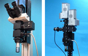 UFS-500
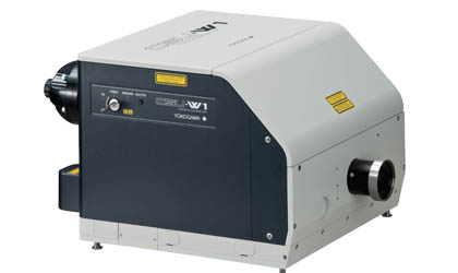
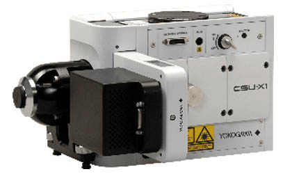
Confocal scanner CSU-W1 CSU-X1
There are wide-field, high-definition models and ultra-high-speed models that enable live-cell observation with high-speed low color fading.
CSU-W1
Wide-field, high-definition model
CSU-X1
ultra high speed model
Focus scanner FS-100
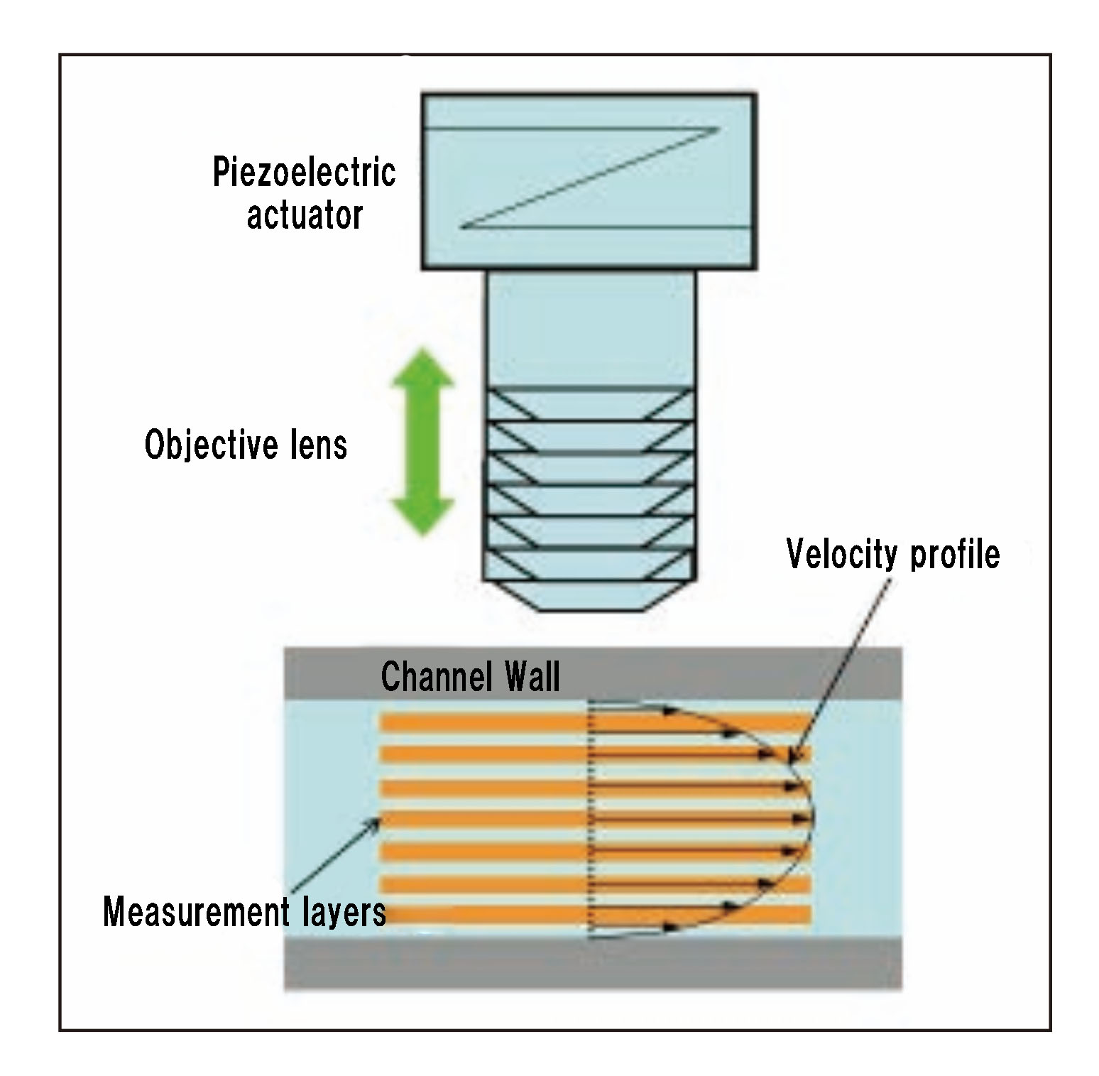
A high-speed microimaging piezofocus scanner suitable for use with high-speed video and confocal scanners. It is installed between the microscope body and the objective lens. This unit scans the observation surface at high speed by moving the objective lens up and down at high speed.
Measurement example
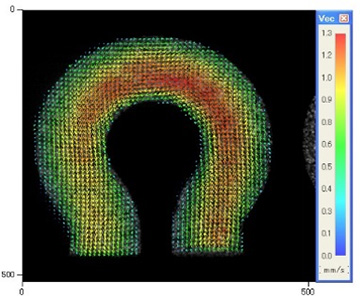
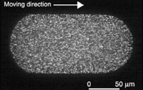
Measurement of 2D-PIV microdroplet internal flow
■ Precise measurement by dividing the inside of a capsule-shaped microdroplet moving in a microchannel with a width of 100μm and a depth of 100μm into 6 sections.
■ Confocal scanning micro PIV system
■ Data provided by: Oshima Laboratory, Institute of Industrial Science, University of Tokyo
Micro PIV measurement example of cooling water flow path
■ Confirmation of cooling efficiency
■ For checking air-cooling fan/water-cooling flow paths
■ Supports millimeter scale/micro scale
■ Compatible with special optics/optical microscopes